
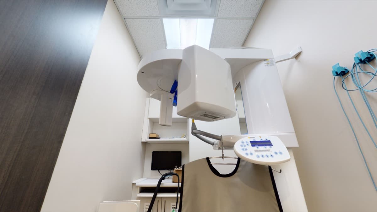
What Are Digital Dental X-Rays?
Digital dental X-rays, also known as digital radiographs, are a modern form of dental imaging that uses electronic sensors instead of traditional photographic film to capture images of your teeth, jawbone, and surrounding structures. These images are displayed instantly on a computer screen, providing your dentist with clear, detailed views that support accurate diagnosis and treatment planning.
At Orion Dental, we use digital X-ray technology at both our Scarborough and Milton offices as a standard part of our diagnostic process. Digital radiography represents a significant advancement over traditional film-based X-rays, offering numerous benefits for both patients and practitioners.
Lower
Radiation dose than film
Instant
Image results
Higher
Resolution than film
Eco
No chemical processing
Radiation Safety at Orion Dental
What Are the Advantages of Digital X-Rays?
Digital X-ray technology offers several meaningful advantages over traditional film radiography. Understanding these benefits can help you appreciate why digital imaging has become an integral part of modern dental care.
Reduced Radiation Exposure
One of the most useful advantages of digital X-rays is the reduction in radiation exposure. Digital sensors need substantially less radiation than traditional film to produce a clear image. The radiation from traditional dental X-rays was already very low, and this further reduction is particularly welcome for patients who require frequent imaging, as well as for children, who are more sensitive to radiation.
Immediate Image Availability
Unlike traditional film X-rays, which require chemical processing in a darkroom, digital X-ray images appear on the computer screen within seconds of being captured. This immediate availability eliminates waiting time and allows your dentist to review your images with you during your appointment, facilitating more efficient consultations and quicker treatment decisions.
Enhanced Image Quality
Digital X-ray images can be enlarged, brightened, darkened, and enhanced using computer software, allowing your dentist to examine specific areas in greater detail. This capability can reveal subtle changes that might be difficult to detect on a traditional film X-ray. The ability to adjust contrast and magnification helps improve diagnostic accuracy without the need for additional X-rays.
Environmentally Responsible
Digital radiography eliminates the need for chemical processing solutions, film packets, and other materials associated with traditional X-rays. This reduces the environmental impact of dental imaging and aligns with our commitment to responsible dental practice.
Easy Storage and Sharing
Digital X-ray images are stored electronically in your patient file, making them easy to access, compare over time, and share securely with other dental professionals or insurance providers when needed. This digital storage also eliminates the risk of lost or damaged film.
What Can a Digital Dental X-Ray Show?
Digital X-rays allow your dentist to detect conditions that are completely invisible to the naked eye. Here is what they commonly uncover:
Cavities Between Teeth
Interproximal decay that forms in the contact areas between teeth — impossible to spot during a visual exam alone.
Bone Loss
Early and advanced bone loss associated with gum disease, helping guide periodontal treatment before damage becomes severe.
Infections at Root Tips
Periapical abscesses and infections at the root apex that cause pain and can spread if left untreated.
Impacted Teeth
Teeth that are blocked from erupting normally — most commonly wisdom teeth — allowing for proactive planning.
Cysts or Tumours
Abnormal growths in the jaw bone or soft tissues, including benign cysts and, rarely, early-stage oral tumours.
Developmental Issues
Missing adult teeth, extra teeth, or abnormal root formation in children and adolescents that benefits from early intervention.
What Types of Dental X-Ray Are There?
At Orion Dental, we use several types of digital X-rays depending on the diagnostic information needed. Your dentist will recommend the type of imaging that is most appropriate for your specific situation.
Bitewing X-Rays
Bitewing X-rays capture the upper and lower teeth in a specific area of the mouth, showing the crowns of the teeth and the bone level between them. They are the most commonly taken dental X-rays and are primarily used to detect cavities between teeth and to monitor bone density changes that may indicate gum disease.
Periapical X-Rays
Periapical X-rays show the entire tooth — from the crown to the tip of the root and the surrounding bone. They are particularly useful for diagnosing issues below the gum line, such as root infections, abscesses, cysts, and bone loss. These X-rays are often taken when a specific tooth is causing symptoms.
Panoramic X-Rays
A panoramic X-ray captures a broad view of the entire mouth in a single image, including all the teeth, the upper and lower jaws, the temporomandibular joints (TMJs), and the sinuses. This type of X-ray is useful for assessing overall oral health, evaluating wisdom teeth, planning orthodontic treatment, and detecting jaw abnormalities.
What Happens During a Dental X-Ray?
Having digital X-rays taken is a quick, comfortable process with no special preparation required. Here is what happens step by step:
Preparation and Shielding
Our team places a lead apron and thyroid collar over you before any imaging begins. This takes just a few seconds and provides an additional layer of protection for areas not being imaged.
Sensor Placement
A small, smooth digital sensor is positioned inside your mouth at the area of interest. It is similar in size to traditional film but has a gentle, rounded edge for comfort.
Image Capture
The X-ray is taken in a fraction of a second. You may notice the sensor in your mouth, and we work to keep you comfortable while it is in place.
Instant Review
Your image appears on the chairside monitor within seconds. Your dentist will walk you through what they see in plain language, pointing out any areas of concern.
Treatment Planning
Based on your X-ray findings, your dentist will discuss any recommended treatments or confirm that everything looks healthy. All images are saved to your file for future comparison.
Ready to experience comfortable, low-radiation digital imaging? Book a checkup at our Scarborough or Milton office today.
Book an AppointmentWhen Are Digital X-Rays Needed?
Digital X-rays play an important role in many aspects of dental care. Your dentist may recommend X-rays in the following situations:
- During routine dental checkups to monitor for cavities, bone loss, and other changes
- Before starting treatment for conditions such as tooth decay, gum disease, or infection
- When planning restorative procedures such as dental implants, crowns, or bridges
- To evaluate the development and positioning of teeth in children and adolescents
- When assessing wisdom teeth to determine whether extraction is necessary
- To investigate the cause of dental pain, swelling, or other symptoms
- Before and after root canal therapy to assess the health of the tooth and surrounding bone
How Much Radiation Does a Dental X-Ray Involve?
Patient safety is a priority at Orion Dental, and our use of digital X-ray technology reflects this commitment. In addition to the inherent radiation reduction offered by digital sensors, we follow the ALARA principle (As Low As Reasonably Achievable) to minimise radiation exposure further. This means that we only take X-rays when there is a clear diagnostic benefit, and we use appropriate shielding, including lead aprons and thyroid collars, to protect areas of the body that are not being imaged.
For children, we take particular care to limit X-ray exposure and use paediatric-sized sensors and settings appropriate for their smaller anatomy. Our team will always explain why a particular X-ray is recommended and answer any questions you may have about the process.
Insurance Coverage for Digital X-Rays
Digital X-Rays and Your Dental Visit
Having digital X-rays taken is a quick and comfortable process. The digital sensor is placed inside your mouth, and the image is captured in just a fraction of a second. There is no discomfort involved, and the results are available immediately for your dentist to review. Your dentist will share the images with you on a chairside monitor and explain what they show, helping you understand your oral health status and any recommended treatments.
If you have been referred to Orion Dental by another dental professional, we can also accept digital X-rays taken at other offices, which can reduce the need for duplicate imaging. Similarly, if you need to be referred to another provider, we can easily share your digital images electronically.
Combining Digital X-Rays with Other Diagnostic Tools
Digital X-rays are often used alongside other modern diagnostic tools to provide the most comprehensive assessment possible. For example, intraoral cameras allow your dentist to capture high-resolution photographs of the visible surfaces of your teeth and gums, complementing the sub-surface views provided by X-rays. Together, these technologies give your dental team a complete picture of your oral health.
Experience Digital X-Rays at Orion Dental
At Orion Dental, we are committed to providing thorough, comfortable dental care using modern diagnostic tools. Digital X-rays are an important part of how we assess and monitor your oral health at both our Scarborough and Milton locations.
To schedule your next appointment, book an appointment online or call our Scarborough office at 416-291-0306 or our Milton office at 905-636-9770. We welcome patients of all ages and accept most major dental insurance plans.